A Case of Streptococcus pneumoniae associated Hemolytic Uremic Syndrome with DIC
Article information
Abstract
Streptococcus pneumoniae associated hemolytic uremic syndrome (SpHUS) is one of the causes of atypical hemolytic uremic syndrome, and increasingly reported. They are more severe and leave more long-term sequelae than more prevalent, typical hemolytic uremic syndrome. But it is not so easy to diagnose SpHUS for several reasons (below), and there was no diagnostic criteria of consensus. A 18 month-old-girl with sudden onset of oliguria and generalized edema was admitted through the emergency room. She had pneumonia with pleural effusion and laboratory findings of HUS, DIC, and positive direct Coombs’ test. As DIC or SpHUS was suspected, we started to treat her with broad spectrum antibiotics, transfusion of washed RBC and replacement of antithrombin III. On the 3rd day, due to severe hyperkalemia and metabolic acidosis, continuous renal replacement therapy (CRRT) was started. She showed gradual improvement in 4 days on CRRT and discharged in 16 days of hospital care. At the follow up to one year, she has maintained normal renal function without proteinuria and hypertension. We report this case with review of articles including recently suggested diagnostic criteria of SpHUS.
Introduction
Hemolytic uremic syndrome is composed of microangiopathic hemolytic anemia, thrombocytopenia and acute renal failure.
Shigatoxin producing E. coli (STEC) cause typical HUS in children. Streptococcus pneumoniae associated HUS (SpHUS) is one of causes of atypical HUS. It is not clear that the increasing incidence of SpHUS is due to the real epidemiological change or improved awareness of the disease. The diagnosis of SpHUS is a little complicated because there is no confirmatory laboratory test, and overlapped clinical and laboratory findings with DIC [1-3]. The mortality and long-term morbidity rates of SpHUS are known to be higher than STEC HUS [4]. Since the introduction of Prevnar 7, the serotypes associated with SpHUS have been changing. With introduction of new vaccines of multivalents (Prevnar 13), we expect a change of epidemiology of SpHUS in near future.
There has been three case reports of SpHUS in Korean children so far [5]. Here we report our case which was complicated with DIC and defined by recently suggested diagnostic criteria including direct Coombs' test (+) to extend our recognition of this rare disease [6].
Case report
A 18-month-old girl was admitted to ER with oliguria and generalized edema for 1 day. She had URI symptoms for 1 week which gradually worsened with fever. One day before admission, generalized edema and weight gain (1.3 kg) was noticed, and on the day of admission generalized seizure with fever developed. At previous hospital, anemia, thrombocytopenia and elevated BUN/Cr were found.
On physical examination at our hospital, she had fever of 38.7℃, blood pressure of 110/75 mmHg, heart rate of 100 beats/minute, respiratory rates of 29/min. She looked acutely illed, mentally alert, anemic and anicteric, but there was no sign of dehydration.
Her lung sounds was decreased on both lung fields and heartbeats were regular without murmur. There was no pitting edema, but mild hepatosplenomegaly (1.5FB).
Her initial laboratory findings revealed hemoglobin of 7.4 g/dL, total leukocyte count of 24,600/mm3, platelet count of 96,000/mm3, ESR of 40 mm/hr and CRP of 3.2 mg/dL.
Urinalysis showed proteinuria (2+) and RBC>30. Peripheral blood smear showed finding of microangiopathic hemolytic anemia (schistocytosis)
Other laboratory results were AST/ALT, 311/101 U/L; LDH, 6419 IU/L; haptoglobin, 20 mg/dL (30-200); BUN/Cr, 48/1.97 mg/dL; PT/aPTT, 1.36/61.6 (24-36 sec.); fibrinogen 133.1 mg/dL (200-400); d-dimer >10.0 mg/dL (0.1-0.85); direct Coombs' test (+); ADAMTS 13 activity, 15% (mild to moderate deficiency).
Her chest radiography and CT demonstrated multifocal consolidations in upper and lower lobes of both lungs with bilateral pleural effusion and cardiomegaly (Fig. 1). Ultrasonography of both kidneys showed enlargement with diffuse increased cortical echogenicity with increased RI value ranged 0.88 to 0.9 (Fig. 2).

Chest CT demonstrates multifocal consolidations in upper and lower lobes of both lungs with bilateral pleural effusion and cardiomegaly.
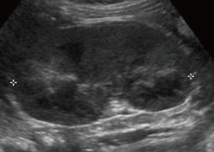
Renal doppler ultrasonography shows enlargement of both kidneys with diffuse increased cortical echogenicity and increased RI value ranged 0.88 to 0.9.
As she was suspected to have DIC or SpHUS, we started to treat her with vancomycin, cefotaxime, transfusion of washed RBC and replacement of antithrombin III. On the 3rd day, due to severe hyperkalemia and metabolic acidosis, continuous renal replacement therapy (CRRT) was started. After 4 days on CRRT, her renal function became to improve, with improvement of other laboratory tests. After 16 days of hospital admission, she was discharged with full recovery. At follow up visits to 1 year after discharge, she has maintained normal renal function without proteinuria and hypertension.
Discussion
Streptococcus pneumoniae associated HUS has been reported in 5-15% of all childhood HUS cases and in 40% of non-STEC cases [3, 7]. The incidence of SpHUS after invasive S. pneumoniae infection is known to be 0.4-0.6% [8].
The global epidemiology of SpHUS is different according to the countries, for example, SpHUS is more prevalent than STEC HUS in Taiwan [4, 7]. It is unclear whether this is due to the rarity of STEC HUS in Taiwan, genetic factor or uncommon use of the pneumococcal vaccine.
In the pre-pneumococcal vaccine era, the prominent serotypes of SpHUS were 14, 6B, 9V, 19, 3, 8 and 23F [3]. After the advent of seven-valents pneumococcal protein conjugate vaccine (Prevnar) in 2000, there was dramatic shift of SpHUS associated strains which were not covered by the vaccine. Newly developed 13-valent vaccine (Prevnar 13) and 23-valent vaccine (Pneumovax 23) now protect against most of strains of SpHUS , but it remains to be seen whether another shift of strains of SpHUS occur. But unluckily, Pneumovax 23 is not recommended in children less than 2 years of age, because it does not develop adequate immune responses in them.
Children under 2 years have the highest incidence of SpHUS, because invasive pneumococcal infections frequently occur to them.
SpHUS were associated with pneumonia usually with pleural effusion or empyema in 62-92% of patient [2, 4, 9].
Meningitis was the second most common condition associated with SpHUS with a higher mortality rate [3]. Spontaneous bacteremia, peritonitis, pericarditis and mastoiditis were rare causes of SpHUS.
The exact pathogenesis is unknown so far, but most accepted theory is that associated with Thomsen-Friedenreich antigen. This normally hidden antigen exists on the surface of RBC, Platelets, glomerular cells and hepatocytes. S. pneumonia can produce neuraminidase, which can expose Thomsen-Friedenreich antigen by cleaving N-acetylneuraminic acid from glycoprotein on the surface of cell membranes. Once the Thomsen-Friedenreich antigen is exposed, it can interact with preformed IgM antibodies. This result in polyagglutination, hemolysis and endothelial injury, then HUS phenotype [10].
More recently Factor H involvement as pathogenesis is suggested. The theory is that Factor H binding sites can be disrupted by desialylation by neuraminidase. As results, Factor H can not bind properly to C3 convertase on the cell surface, leading to complement activation and cell injury [11].
A certain serotype (serotype 3) of pneumococcus express Hic protein which bind Factor H directly [12].
SpHUS typically develops between 3 to 13 days after pneumococcal infection [3]. Compared to STEC HUS, patients with SpHUS tend to be younger, have more severe renal, hematologic and neurological disease (longer duration of oliguria and dialysis, more transfusion, longer period of thrombocytopenia) [3].
Extra-renal complications are frequent including pancreatitis, purpura fulminans, extremity amputation, cholecystitis, thrombosis and hearing loss [9]. Other rare findings are hemidiaphragmatic paralysis, hepatosplenomegaly, reticulocytopenia and hepatitis.
Overall, SpHUS has higher mortality and long-term morbidity rates than STEC HUS. In review of all known cases of SpHUS reported in the English language literature until 2008, 12% died, 10% progressed to end-stage kidney disease, and 16% survived with chronic kidney disease or hypertension [3]. Long-term outcomes for SpHUS are difficult to ascertain because of limited studies and short follow-up periods. The long-term clinical significance of mild increases in serum creatinine concentration or mild proteinuria is unknown. The risk factor for the development of chronic kidney is the need for acute dialysis for more than 20 days.
The diagnosis of SpHUS might be difficult in critically ill patients due to the similarities in laboratory and clinical findings in DIC. In addition, both conditions might coexist in a child with pneumococcal infection. Marked prolongation of coagulation time, increased FDP and decreased serum fibrinogen titer help to distinguish between SpHUS and DIC. Second reason in difficulty in the diagnosis is the bacteriological confirmation is not so easy because the sputum culture from baby less than 2 yeas of age is very difficult. Thirdly, there is no specific laboratory tests to the diagnosis of SpHUS.
The direct Coombs’ test detects antibodies and complement that coat RBC and can be used as a screening test for SpHUS [13]. The positive Coombs’ test is very sensitive for SpHUS, but the specificity is unclear.
The peanut lectin agglutination method can detect Thomsen-Friedenreich antigen exposure. In 36 patients with invasive pneumococcal disease, the peanut lectin activity assay showed high sensitivity (100%), but low specificity(48%) for SpHUS. Canadian Pediatric Society defines definitive cases as evidence of S. pneumoniae infection, evidence of HUS and presence of thrombotic microangiopathy on biopsy.
But their criteria for the diagnosis of SpHUS is so strict that many cases of SpHUS might be missed and lose opportunities to be adequately treated.
Recently, based on a combination of clinical and laboratory findings and evidence of S. pneumoniae infection, a modified definition for SpHUS was proposed by Copelovitch that divided cases into three categories: definite, probable or possible [6] (Table l). According to that definition, our case belongs to possible category.

Modified Pneumococcal Hemolytic Uremic Syndrome Case Definitions
The treatment of SpHUS is mainly supportive like STEC HUS, that include fluid and electrolyte balance. Dialysis or CRRT is often required for prolonged time.
As initial treatment of invasive pneumococcal infection in critically ill children, addition of vancomycin to extended spectrum cephalosporin could be considered [14]. As soon as the sensitivity tests demonstrate effectiveness of other antibiotics, vancomycin should be discontinued. Due to the concern for preformed anti-Thomsen-Friedenreich antibodies, use of washed RBC when needed and avoidance of fresh frozen plasma [FFP] use are important.
In summary, our case of 18-month-old female with pneumonia and DIC was diagnosed with SpHUS by newly developed diagnostic criteria including positive Coombs’ test result and treated with broad spectrum antibiotics, transfusion of washed RBC and CRRT with complete recovery. We think it is important to diagnose SpHUS expeditiously by new criteria of diagnosis and treat promptly with adequate antibiotics, avoiding use of fresh frozen plasma.
Notes
Conflict of interest
No potential conflict of interest relevant to this was reported.
Acknowledgements
This study was supported for 2 years by research grant from Pusan National University